Prokaryotic (Bacterial) Cell Model: 3D Parts, Gram Stain & Antibiotics Lab
🦠 Interactive 3D Bacterium
Interactive 3D model of an E. coli bacterium drawn to true proportions, about 2 micrometres long. It is wrapped in a capsule, an outer membrane, a thin peptidoglycan cell wall and the cell membrane. Inside, a long folded loop of DNA forms the nucleoid, with small plasmid rings, storage granules and thousands of ribosomes in the crowded cytoplasm. Several corkscrew-shaped flagella, spun by motors in the cell envelope, bundle behind the cell, and hundreds of short fimbriae and one long sex pilus stick out from the surface. Drag or use the arrow keys to turn it, plus and minus to zoom, and tap a part to read about it.
Or press “Take the tour”. Try “Cut open” to see the layers and the DNA inside.
Shape
Cell wall type
Find the parts 0 / 9
Drawn to true proportions for E. coli (about 2 µm long and 0.9 µm wide). The thin layers, the DNA and the pili are drawn several times thicker so you can see them, the flagella are shortened, and a real cell holds even more ribosomes. Swimming is shown slowed down: a real E. coli swims about 12 body lengths per second.
🎓 For teachers: a 40-minute lesson with this page
- Hook (5 min): project the page in full screen (⛶) and press Take the tour.
- Explore (10 min): students find all 9 parts and do the Label quiz below. Ask: what is missing compared with an animal cell?
- Gram stain lab (10 min): stain each sample, then identify two mystery samples. Discuss why over-decolorizing gives a false result.
- Antibiotics (10 min): run the dish with 0 % and then 5 % resistant cells. Ask: did the drug make the resistant cells?
- Exit ticket (5 min): the final quiz (10 questions). Missions are saved in each student’s browser; finishing all 8 unlocks a printable certificate.
Works in Google Classroom, Nearpod (Web Content) and Teams: just share the link. No sign-up, no ads, works on Chromebooks and phones.
📏 Mission 2 · How Small Is a Bacterium?
Zoom from a grain of salt down to the width of DNA. Everything is drawn to the same scale. Tap an object to zoom to it.
🧪 Mission 3 · Gram Stain Virtual Lab
The Gram stain is the first test a microbiologist does on an unknown bacterium. It sorts bacteria into two groups by their cell wall. Stain a slide step by step, predict the color, then look under the microscope. When you are ready, identify a mystery sample.
What is happening in the cell wall:
In a real lab: heat-fix the smear, then crystal violet about 60 s, iodine about 60 s, decolorizer (ethanol or acetone–alcohol) for just a few seconds, safranin about 60 s, rinsing with water between steps. Times vary by lab; this is a simplified teaching model.
➗ Mission 4 · Binary Fission & Growth
How one bacterium becomes two
Doubling, doubling, doubling…
Grow a culture in a test tube
Real growth does not go on forever. Pick a temperature and watch 24 hours of growth (start: 1,000 cells per mL).
·
Teaching model based on E. coli: fastest growth (about 20 min per doubling) near body temperature, slow growth at room temperature, almost none in the fridge, and death when heated. The four phases are lag, log, stationary and death.
💊 Mission 5 · Antibiotics & Resistance Lab
A dish of E. coli. Some cells (orange) carry a plasmid with a gene that destroys ampicillin. Choose how many start resistant, add an antibiotic, and run 8 hours. Try: 0 % resistant + ampicillin, then 5 % + ampicillin.
Time 0 h 00 min · ● sensitive 0 · ● resistant 0
Ampicillin stops the enzymes that cross-link the peptidoglycan wall. A growing cell gets weak spots and bursts. Our cells have no peptidoglycan, so they are not harmed. The resistance plasmid’s gene makes an enzyme (β-lactamase) that breaks the drug apart.
Tetracycline sticks to the small (30S) part of the bacterial ribosome, so no new tRNAs can bring amino acids. With no new proteins, the cells stop growing (the drug is bacteriostatic). In this lab the plasmid gives no protection against it.
Teaching model. Real resistance plasmids, rates and doses vary. Always take antibiotics exactly as a doctor or pharmacist tells you, and never use them for colds or flu: those are caused by viruses.
🏊 Mission 6 · Swim to Food (Chemotaxis)
E. coli cannot see or steer. It swims straight (a run), then its flagella fly apart and it tumbles to a random new direction. Its only trick: when the sugar is getting stronger, it waits longer before tumbling. Can you reach the sugar using only the bacterium’s sense?
🏷️ Mission 7 · Labeled Bacterial Cell Diagram, Label Quiz & Functions
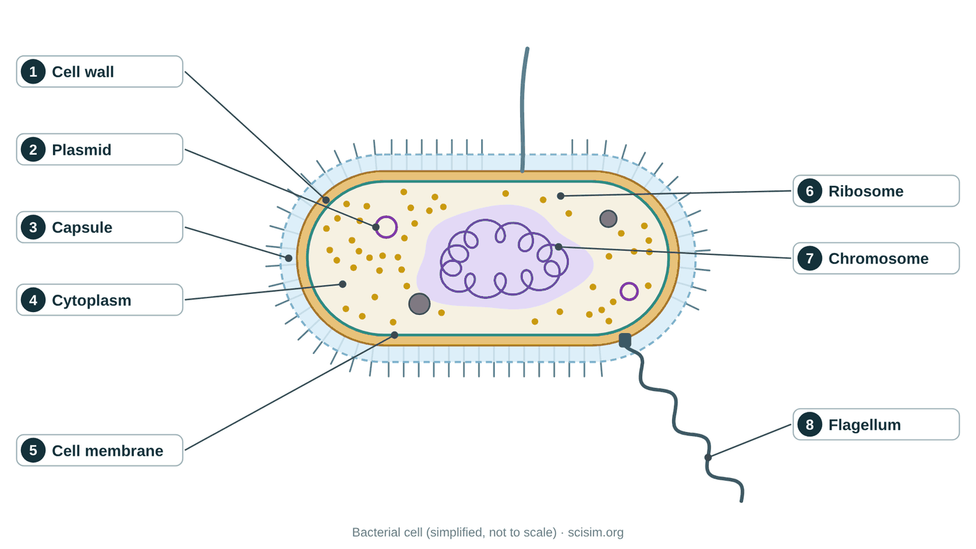
The 3D model above shows the cell in depth; this flat diagram is the version you draw in an exam. Pick Basic (the 8 parts most courses ask for) or Advanced (12 parts). Then switch between the labeled diagram, a blank one to test your memory, a label quiz where you place each label yourself, and match the jobs, where you pair each part with what it does. Not every bacterium has a capsule, flagellum, pili or plasmids. You can also download the diagrams or print a worksheet with an answer key. On a phone, swipe the diagram sideways.
Tap a word, then tap the numbered box it belongs to (on the diagram or in the list below). Tap a filled box to take the word back.
Each line describes the job of one part. Tap a part name, then tap the box next to the job it does. Tap a filled box to take the word back.
✅ Mission 8 · Final Quiz
10 questions from a bank of 20. Get 6 or more to complete the mission. Press “New quiz” at the end for different questions.
💡 The Idea, Step by Step
Bacteria were the first life on Earth, and they are still everywhere — in soil, in the ocean, and trillions of them living in and on you. A bacterium is a prokaryotic cell, from Greek for "before the nut (kernel)," meaning before the nucleus. It is tiny: a typical rod is only about $1$–$5\,\mu\mathrm{m}$ long, roughly ten times smaller across than an animal cell. Everything it needs to live is packed into one simple compartment.
The defining feature is what a bacterium lacks: there is no nucleus and no membrane-bound organelles — no mitochondria, no ER, no Golgi. Instead the main DNA, a single circular chromosome, sits loose in a region called the nucleoid. Floating nearby are small DNA rings called plasmids and huge numbers of ribosomes making proteins. Around the cell are a plasma membrane, a rigid cell wall of peptidoglycan, and often a slimy capsule. Many bacteria also have a flagellum for swimming and pili for sticking and sharing DNA.
"Simple" does not mean weak. Because there is no nuclear membrane separating the DNA, a bacterium can transcribe a gene into mRNA and translate it into protein at the same time, so it responds to its environment almost instantly. Its small size gives it a large surface-area-to-volume ratio, $\frac{A}{V}$, so nutrients reach every part fast. It can divide as often as every $20$ minutes, and share plasmid genes (including antibiotic resistance) with its neighbours. The cell wall is so important — it stops the cell bursting under osmotic pressure — that antibiotics like penicillin kill bacteria precisely by attacking it.
Press Cut open and find the nucleoid: the DNA is loose, not boxed in a nucleus. Switch between Gram − and Gram + and compare the walls, then run the same two kinds of bacteria through the Gram stain lab. In the antibiotics lab, start with 0 % and then 5 % resistant cells: the drug kills the sensitive cells but cannot create resistance. Finally, set the test tube to 4 °C and then 37 °C to see why we keep food in the fridge.
📐 The Parts of a Bacterium
| Part | Main job | In eukaryotes too? |
|---|---|---|
| Capsule | Sticky outer slime layer; protection and sticking to surfaces | No (some have a coat) |
| Cell wall | Rigid peptidoglycan layer; shape and stops bursting | Plants/fungi have walls (different material) |
| Plasma membrane | Selective barrier; also does jobs the membrane organelles do in eukaryotes | Yes |
| Cytoplasm | Watery interior where reactions happen | Yes |
| Nucleoid | Region holding the single circular chromosome (main DNA) | No (eukaryotes use a nucleus) |
| Plasmid | Small extra DNA ring with bonus genes (e.g. resistance) | Rare |
| Ribosomes | Build proteins; smaller (70S) than eukaryotic (80S) | Yes (larger) |
| Flagellum | Whip-like tail spun by a motor for swimming | Different kind exists |
| Pili / fimbriae | Hair-like surface fibres for attaching and transferring DNA | No |
The membrane does the organelles' work
Since a bacterium has no mitochondria or chloroplasts, the reactions those organelles perform in eukaryotes happen on the plasma membrane instead. The proteins of the electron transport chain that make ATP sit right in the bacterial membrane, and in photosynthetic bacteria the light reactions happen on infolded membrane patches. One simple boundary does many of the jobs that eukaryotes spread across separate compartments — an elegant consequence of being small.
Why ribosome size matters for medicine
Bacterial ribosomes (70S) are built slightly differently from our own (80S). Several antibiotics — such as tetracyclines and erythromycin — exploit this difference, jamming the bacterial ribosome while leaving human ribosomes untouched. This idea, called selective toxicity, is why we can kill bacteria inside our bodies without poisoning ourselves, and it is a direct, practical payoff of understanding prokaryotic cell structure.
🎓 Going Further (AP / A-level / Intro College)
Gram-positive vs Gram-negative envelopes
| Feature | Gram-positive (e.g. Staphylococcus, Bacillus) | Gram-negative (e.g. E. coli) |
|---|---|---|
| Peptidoglycan | Thick, many layers (about 20–80 nm), with teichoic acids | Thin, one or a few layers (about 7 nm), in the periplasm |
| Outer membrane | None | Yes, outer leaflet of lipopolysaccharide (LPS); porins let small molecules through |
| Gram stain | Keeps crystal violet–iodine → purple | Loses it after decolorizing → pink with safranin |
| Penicillin G / lysozyme | Usually more sensitive | Outer membrane blocks many drugs; LPS (endotoxin) can trigger fever and shock |
The growth equation
In the log phase each cell divides after a doubling time $g$, so after time $t$ a population of $N_0$ becomes $N = N_0 \cdot 2^{t/g}$. The number of generations is $n = t/g$, and the growth rate constant is $k = \ln 2 / g$. Example: $N_0 = 10^3$ cells/mL, $g = 20$ min, $t = 4$ h gives $n = 12$ and $N = 10^3 \times 4096 \approx 4.1 \times 10^6$ cells/mL. Real cultures leave the log phase when nutrients run out and waste builds up (stationary phase).
How antibiotics are selectively toxic
| Target | Example drugs | Why our cells are spared |
|---|---|---|
| Cell wall synthesis | Penicillins (ampicillin), cephalosporins, vancomycin | Human cells have no peptidoglycan |
| 70S ribosome | Tetracyclines (30S), macrolides such as erythromycin (50S), aminoglycosides (30S) | Our cytoplasmic ribosomes are 80S |
| DNA replication | Fluoroquinolones (block DNA gyrase) | Our topoisomerases are different |
| Folate synthesis | Sulfonamides; trimethoprim | Sulfonamides: we do not make folate (we get it from food). Trimethoprim: our version of the target enzyme (DHFR) is different |
Three ways bacteria share genes (horizontal gene transfer)
Conjugation: a donor with a conjugative plasmid makes a sex pilus, pulls a recipient close and passes a single strand of the plasmid, which both cells copy. Transformation: a cell takes up free DNA from its surroundings (Griffith’s experiment). Transduction: a bacteriophage carries bacterial DNA from one cell to another. Together with fast growth, this is why resistance can spread through a population quickly.
Physics of being small
For a bacterium, water is like thick syrup: the Reynolds number is about $10^{-5}$, so there is no gliding. If the flagellar motor stops, the cell stops in about a microsecond, after coasting less than the width of an atom (Purcell, “Life at Low Reynolds Number”, 1977). That is why bacteria use a rotating corkscrew, which keeps pushing, instead of a flapping tail. The motor is driven by protons flowing into the cell and spins at around 100 (up to about 300) revolutions per second (Berg, E. coli in Motion, 2004).
❓ FAQ
A prokaryotic cell is a cell with no membrane-bound nucleus and no membrane-bound organelles. Bacteria and archaea are prokaryotes. Their DNA floats freely in a region of the cytoplasm called the nucleoid rather than being enclosed in a nucleus. Prokaryotic cells are also much smaller and simpler than eukaryotic cells.
Key takeaway: a prokaryote keeps its DNA loose in the cytoplasm and has no membrane-bound nucleus or organelles.A bacterium's main DNA is a single, usually circular chromosome that sits in an irregular region of the cytoplasm called the nucleoid. It is not wrapped in a membrane, so it is in direct contact with the rest of the cell. Many bacteria also carry small extra rings of DNA called plasmids.
Key takeaway: bacterial DNA is a circular chromosome in the nucleoid, with no surrounding nuclear membrane.A plasmid is a small, circular piece of DNA separate from the main chromosome. It carries a few extra genes — often ones for antibiotic resistance or for breaking down unusual nutrients — that are useful but not essential. Bacteria can copy plasmids and pass them to other bacteria, which is a major way antibiotic resistance spreads. Plasmids are also the workhorses of genetic engineering.
Key takeaway: a plasmid is a small extra DNA ring carrying bonus genes that bacteria can share, including resistance.The bacterial cell wall is a rigid layer outside the plasma membrane, made mainly of peptidoglycan. It gives the cell its shape and, crucially, stops it from bursting when water flows in by osmosis. Because peptidoglycan is unique to bacteria, many antibiotics such as penicillin work by attacking the cell wall without harming our own cells.
Key takeaway: the peptidoglycan cell wall gives shape and prevents bursting, and is a key antibiotic target.Many bacteria swim using a flagellum, a long whip-like tail driven by a tiny rotary motor embedded in the membrane and wall. The motor spins the flagellum like a propeller, pushing the cell through liquid. Bacteria steer toward food or away from danger by changing how the motor turns, a behaviour called chemotaxis.
Key takeaway: bacteria swim by spinning a flagellum with a rotary motor, steering by chemotaxis.Prokaryotic cells (bacteria, archaea) have no nucleus and no membrane-bound organelles, their DNA is a single circular chromosome in the nucleoid, and they are small (about $1$–$5\,\mu\mathrm{m}$) with small (70S) ribosomes. Eukaryotic cells have a true nucleus, many membrane-bound organelles, usually linear chromosomes, and are larger with larger (80S) ribosomes. Both have a plasma membrane, cytoplasm, ribosomes, and DNA.
Key takeaway: prokaryotes are small and lack a nucleus and organelles; eukaryotes are larger and compartmentalised — but both share membranes, cytoplasm, ribosomes, and DNA.Being small and simple is an advantage. Bacteria reproduce extremely fast (some divide every $20$ minutes), so they evolve quickly and adapt to new environments. They can share genes directly through plasmids, exploit almost every habitat on Earth, and form protective communities called biofilms. Their lack of a nucleus actually lets transcription and translation happen at the same time, speeding up protein production.
Key takeaway: fast reproduction, gene sharing, and metabolic flexibility make "simple" prokaryotes one of the most successful forms of life on Earth.The Gram stain sorts bacteria into two groups by their cell wall. Cells are stained with crystal violet, then iodine is added as a mordant that locks the dye in, then they are washed briefly with alcohol or acetone and counterstained with safranin. Gram-positive bacteria have a thick peptidoglycan wall that traps the purple dye, so they stay purple. Gram-negative bacteria have a thin wall plus an outer membrane, lose the purple and look pink. Doctors use the result, together with the cell shape, as a fast first clue to which antibiotics may work.
Key takeaway: Gram-positive = thick wall = purple; Gram-negative = thin wall + outer membrane = pink.Bacteria reproduce by binary fission: they copy their DNA, grow and split into two. In ideal conditions E. coli can divide about every 20 minutes, so one cell could become 2 to the power 24, about 17 million cells, in 8 hours. In real life growth slows and stops when food runs out and waste builds up (the stationary phase). Cold slows growth a lot, which is why we refrigerate food, and enough heat kills bacteria.
Key takeaway: Doubling every 20 minutes is exponential growth — but food, waste and temperature limit it.Antibiotics do not make bacteria resistant. In a large population a few cells may already carry a resistance gene, often on a plasmid. The antibiotic kills the sensitive cells and the resistant ones survive and multiply — natural selection. Bacteria can also pass resistance plasmids to their neighbours by conjugation through a sex pilus, so resistance can spread even between different species. Using antibiotics only when needed, and exactly as prescribed, slows this down.
Key takeaway: Antibiotics select resistant bacteria that already exist; plasmids then spread the genes.Antibiotics attack parts of bacteria that are missing or different in our cells, such as the peptidoglycan cell wall or the bacterial 70S ribosome. A virus is not a cell: it has no cell wall and no ribosomes of its own — it uses the host cell’s machinery. So an antibiotic has nothing to hit. Colds and flu are caused by viruses, so antibiotics do not help them.
Key takeaway: No wall and no ribosomes of its own means nothing for an antibiotic to attack.⚠️ Misconceptions & Common Errors