Animal Cell Model: 3D Organelles, Microscope & Protein Labs
🔬 Interactive 3D Animal Cell
Interactive 3D model of an animal cell drawn to true proportions, about 20 micrometres across, with a flexible cell membrane and no wall. Inside are a large nucleus with nuclear pores and a nucleolus, rough ER studded with ribosomes, smooth ER tubes, a pink Golgi stack with vesicles, sausage-shaped mitochondria with folded cristae, lysosomes, peroxisomes, thousands of free ribosomes, and a centrosome with microtubules spreading through the cytoplasm. Drag or use the arrow keys to turn it, plus and minus to zoom, and tap a part to read about it.
Or press “Take the tour”. Try “Cut open” to see inside.
Find the parts 0 / 10
Drawn to true proportions for a typical animal cell (about 20 µm across). Membranes, ER, ribosomes and fibers are drawn several times thicker than true so you can see them; a real cell has millions of ribosomes. Cells come in many shapes — this one is a generalized cell.
🎓 For teachers: a 40-minute lesson with this page
- Hook (5 min): project the page in full screen (⛶) and press Take the tour.
- Explore (10 min): students find all 10 parts, then do the protein journey. Ask: why do gland cells have lots of rough ER and Golgi?
- Microscope lab (10 min): stain cheek cells, focus up to 400× and estimate the cell size from the field of view.
- Osmosis (10 min): put a red blood cell in pure water, then in strong salt water. Compare with a plant cell.
- Exit ticket (5 min): the final quiz. Missions are saved in each student’s browser; finishing all 8 unlocks a printable certificate.
Works in Google Classroom and Teams: just share the link. No sign-up, no ads, works on Chromebooks and phones.
🔬 Mission 2 · Microscope Lab: Cheek Cells
Prepare a slide, choose the right stain, add the coverslip without air bubbles and focus up to high power — then measure a cell using the field of view. This is the practical every biology student does.
Objective lens
Coarse focus (big knob)
Fine focus (small knob)
The objective is the lens near the slide. The eyepiece is 10×, so a 4× objective gives 10 × 4 = 40× in total (10× gives 100×, 40× gives 400×). Field-of-view sizes are typical for a school microscope; check your own with a clear ruler at low power.
📦 Mission 3 · A Protein’s Journey
Salivary gland cells make the enzyme amylase and send it out into your mouth. Tap the parts of the cell in the order the protein travels.
💧 Mission 4 · Water In, Water Out (Osmosis)
Water moves by osmosis from a dilute solution to a more concentrated one, through the partly permeable cell membrane. Change the solution outside the cell. Make the red blood cell burst and shrink, then plasmolyse a plant cell (make its membrane pull away from the wall) to see what a cell wall changes.
Teaching model. 0.9% salt (about 300 mOsm per litre) is isotonic with blood; red blood cells begin to burst below about 0.45%. The onion cell sap is taken as about 0.3 M sucrose; real values vary between plants.
⚖️ Mission 5 · Compare Animal, Plant & Bacterial Cells
Tick every cell type that has each feature, then press Check. Some features are in all three.
See the 3D bacterial cell and the 3D plant cell to compare in more detail.
📏 Mission 6 · How Small Is a Cell?
Everything is drawn to the same scale. Tap an object to zoom to it — try the animal cell, then zoom down to a ribosome.
🏷️ Mission 7 · Labeled Animal Cell Diagram & Label Quiz
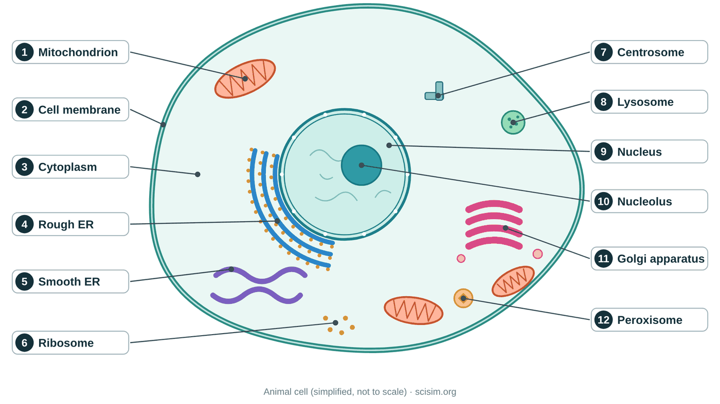
The 3D model above shows the cell in depth; this flat diagram is the version you draw in an exam. Switch between the labeled diagram, a blank one to test your memory, and a quiz where you place each label yourself. You can also download both versions or print a worksheet.
Tap a word, then tap the numbered box it belongs to (on the diagram or in the list below). Tap a filled box to take the word back. On a phone, swipe the diagram sideways to see every number.
✅ Mission 8 · Final Quiz
10 questions from a bank of 20. Get 6 or more to complete the mission.
💡 The Idea, Step by Step
Think of an animal cell as a busy factory sealed inside a soft, water-filled bag. The bag is the cell membrane, and the watery space inside is the cytoplasm. A typical animal cell is only about $10$–$30\,\mu\mathrm{m}$ wide — far too small to see without a microscope — yet inside it dozens of little machines are all working at once. Each machine is an organelle ("little organ"), and just like the organs in your body, every organelle has one main job.
At the center sits the nucleus, the manager: it holds the cell's DNA and the instructions for every protein. Scattered around it are the mitochondria, the power plants that burn fuel to make ATP energy. A folded membrane network called the endoplasmic reticulum (ER) builds proteins (on its ribosome-studded rough region) and lipids (on its smooth region). The Golgi apparatus is the shipping department, and the lysosomes are the recycling crew. Click each one in the sim to meet it.
The big idea is compartmentalization: by wrapping jobs in separate membranes, one cell can run many chemical processes that would interfere with each other if mixed. A lysosome can stay acidic while the cytoplasm stays neutral; a mitochondrion can build a steep proton gradient; the nucleus can keep its DNA safe. Folded membranes also pack in enormous surface area — the ratio $\frac{\text{surface area}}{\text{volume}}$ is what lets a mitochondrion's cristae and the sprawling ER host so many reactions. The parts also form a pathway: nucleus → ribosome/rough ER → Golgi → vesicle → destination.
Press Cut open and find the nucleus, the rough ER wrapped around it and the pink Golgi stack. Then follow the same route in the protein journey. Switch on Bacterium for scale to see how small a whole bacterium is next to this cell. In the osmosis lab, put a red blood cell into pure water: with no cell wall, it bursts.
📐 The Organelles and Their Jobs
| Organelle | Main job | Membrane? |
|---|---|---|
| Cell membrane | Selectively controls what enters and leaves; the cell's boundary | Single bilayer |
| Nucleus | Stores DNA and directs protein synthesis (the control center) | Double (nuclear envelope) |
| Nucleolus | Makes ribosomal RNA and assembles ribosome subunits | None (region in nucleus) |
| Mitochondrion | Aerobic respiration → ATP energy; has its own DNA | Double (inner = cristae) |
| Rough ER | Synthesizes and folds proteins (ribosome-studded) | Single |
| Smooth ER | Makes lipids, stores calcium, detoxifies | Single |
| Golgi apparatus | Modifies, sorts, and ships proteins and lipids in vesicles | Single (stacked sacs) |
| Lysosome | Digests and recycles waste with acidic enzymes | Single |
| Ribosome | Reads mRNA and builds proteins | None |
| Centrosome | Organizes microtubules and the spindle in cell division | None |
| Peroxisome | Breaks down fatty acids; neutralizes hydrogen peroxide | Single |
The protein production line (the endomembrane system)
Many of these organelles are physically connected as one team called the endomembrane system. The nuclear envelope is continuous with the rough ER; a gene is copied into mRNA in the nucleus, that mRNA is read by a ribosome on the rough ER, and the new protein is folded inside the ER. Vesicles then bud off and carry the protein to the Golgi, which tags and sorts it before shipping it onward — to a lysosome, to the membrane, or out of the cell entirely. This is why the rough ER, Golgi, and nucleus sit so close together in the sim.
Why mitochondria have folds
The energy reactions of respiration happen on the inner mitochondrial membrane. By folding that membrane into cristae, the mitochondrion packs far more membrane — and therefore far more of the protein machinery — into the same small volume, boosting ATP output. Mitochondria also carry their own small circular DNA and replicate on their own, evidence for the endosymbiotic theory that they descend from ancient free-living bacteria.
🎓 Going Further (AP / A-level / Intro College)
The endomembrane system
Proteins for export or for membranes start being made on free ribosomes. A short signal peptide at the start of the protein is recognized by a signal-recognition particle, which docks the ribosome onto the rough ER. The growing protein is threaded into the ER, folded and often given sugar groups, then carried in vesicles to the Golgi, modified again and sorted to the membrane, to lysosomes or out of the cell (exocytosis). Proteins without a signal stay in the cytosol.
Why cells are small: surface area to volume
For a cube of side $L$, surface area $= 6L^2$ and volume $= L^3$, so $SA:V = 6/L$. A 10 µm cell has $SA:V = 0.6$ per µm; a 20 µm cell only $0.3$. Doubling the size halves the membrane available for each unit of volume to take in oxygen and nutrients and get rid of waste. That is one reason cells stay small and divide, and why some cells (like those lining the gut) have their membrane folded into microvilli.
Magnification and real size
$\text{magnification} = \dfrac{\text{image size}}{\text{actual size}}$. A nucleus 12 mm wide in a photo taken at ×2,000 is really $12\,000\,\mu m / 2000 = 6\,\mu m$ wide. Convert units first: 1 mm = 1,000 µm and 1 µm = 1,000 nm.
Endosymbiosis
Mitochondria are thought to come from free-living bacteria taken in by an ancestral cell (Margulis, 1967). Evidence: a double membrane, their own circular DNA, 70S ribosomes like bacteria, and division by a process like binary fission.
🔎 How we checked this page
Checked against:
- Campbell & Reece — Biology (Ch. 6, “A Tour of the Cell”)
- Alberts et al. — Molecular Biology of the Cell (Ch. 12–15)
Key numbers: A typical animal cell is about 20 µm across, and the 3D model keeps the parts in true proportion to it. The full list is in the references above.
What the model simplifies: Membranes, the ER, ribosomes and fibers are drawn several times thicker than they really are so you can see them, and a real cell has far more ribosomes.
Standards: NGSS MS-LS1-1, MS-LS1-2
Last reviewed · How we check our science · Report a mistake
❓ FAQ
An organelle is a specialized structure inside a cell that performs a specific job, much like an organ does for the whole body. Most organelles in an animal cell are wrapped in their own membrane — the nucleus, mitochondria, endoplasmic reticulum, Golgi apparatus, and lysosomes are examples. A few important structures, like ribosomes, are not membrane-bound.
Key takeaway: organelles divide the cell's work into specialized, often membrane-enclosed compartments.The nucleus is the control center of the cell. It stores the cell's DNA, and the genes in that DNA direct which proteins the cell makes. A double membrane called the nuclear envelope surrounds it, and pores in that envelope let messenger RNA and other molecules pass in and out. Inside is the nucleolus, where ribosomes are assembled.
Key takeaway: the nucleus protects the DNA and directs the cell by controlling protein synthesis.Mitochondria carry out aerobic cellular respiration: they use oxygen to release the energy stored in glucose and capture it in molecules of ATP, the cell's usable energy currency. Their inner membrane is folded into cristae that increase the surface area for the reactions of the electron transport chain. Cells that need lots of energy, like muscle cells, contain thousands of mitochondria.
Key takeaway: mitochondria make most of the cell's ATP through aerobic respiration.The rough endoplasmic reticulum, studded with ribosomes, makes and folds proteins, while the smooth ER makes lipids. Both pinch off vesicles that carry their cargo to the Golgi apparatus. The Golgi then modifies, sorts, and packages those molecules and ships them in new vesicles to their destinations inside or outside the cell.
Key takeaway: the ER manufactures proteins and lipids, and the Golgi finishes, labels, and ships them — an assembly line.A lysosome is a membrane sac filled with acidic digestive (hydrolytic) enzymes. It breaks down worn-out organelles, food particles brought into the cell, and invading bacteria, then recycles the useful building blocks. Its membrane keeps those powerful enzymes safely separated from the rest of the cell.
Key takeaway: lysosomes are the cell's recycling and waste-disposal centers.Animal and plant cells share most organelles — nucleus, mitochondria, ER, Golgi, and ribosomes. Plant cells additionally have a rigid cell wall outside the membrane, chloroplasts for photosynthesis, and a large central vacuole. Animal cells lack all three, but they have centrioles and typically many small lysosomes.
Key takeaway: plant cells add a cell wall, chloroplasts, and a big vacuole; animal cells rely on centrioles and lysosomes.Membranes let the cell create separate compartments, each with its own chemistry. The acidic interior of a lysosome, the energy-converting space inside a mitochondrion, and the protected DNA of the nucleus all depend on a membrane keeping conditions different from the surrounding cytosol. Membranes also vastly increase usable surface area for reactions, which is why the inner mitochondrial membrane and the ER are so folded.
Key takeaway: membranes create specialized compartments and add reaction surface area, letting one cell run many incompatible processes at once.Gently rub the inside of your cheek with a clean cotton bud, smear it on a slide and add a drop of methylene blue stain. Lower a coverslip slowly at an angle to avoid air bubbles. Start with the 4× objective and the coarse focus knob, then move to 10× and 40× and use only the fine focus. The cells look flat and irregular, and the stain shows each nucleus dark blue.
Key takeaway: Cotton bud smear, methylene blue, careful coverslip, and start at low power.Water moves into the cell by osmosis because the inside of the cell is more concentrated than pure water. Animal cells have no cell wall to resist this, so the red blood cell swells into a sphere and bursts (hemolysis). In a very salty solution the opposite happens: water leaves, and the cell shrinks and crinkles. Blood plasma is about the same concentration as 0.9% salt, so cells keep their shape.
Key takeaway: With no wall, an animal cell can burst in pure water.The gene is copied into mRNA in the nucleus. Ribosomes on the rough ER read it and build the protein, which is folded inside the ER. A vesicle carries it to the Golgi apparatus, which modifies and packs it into another vesicle. That vesicle fuses with the cell membrane and releases the protein outside (exocytosis). Mitochondria supply the ATP energy for these steps.
Key takeaway: Nucleus → rough ER → vesicle → Golgi → vesicle → membrane.Most animal cells are about 10–30 micrometres across — around 50 typical cells in a row would make one millimetre. Some are much bigger (a large fat cell can be about 0.1 mm, just visible to the eye) and some nerve cells have extensions about a metre long. Because they are so small, you need a light microscope to see them and an electron microscope to see their organelles in detail.
Key takeaway: Typical animal cells are about 20 µm — too small to see without a microscope.⚠️ Misconceptions & Common Errors