Human Heart Model: Real 3D Heart, Valves, Blood Flow & Pulse Lab
❤️ Interactive 3D Heart
Interactive 3D model of a real adult human heart from the NIH Human Reference Atlas, seen from the front with the apex at the bottom. The right side (right atrium and right ventricle) is on the viewer’s left. From outside the heart is shown in real tissue colors with yellow fat in its grooves; cut open, the walls keep their real color, the lining of the right side has a faint blue tint for oxygen-poor blood and the left side a faint red tint, and blue and red markers show the blood. The aorta arches over the top, the pulmonary trunk splits into the pulmonary arteries, the venae cavae enter the right atrium and four pulmonary veins enter the left atrium at the back. Coronary arteries and cardiac veins run over the surface. Cut open, the four chambers, the septum, the tricuspid and mitral valves, the papillary muscles and the aortic and pulmonary valves are visible. The heart beats through the cardiac cycle and small markers show blood flowing. Drag or use the arrow keys to turn it, plus and minus to zoom, tap a part to read about it.
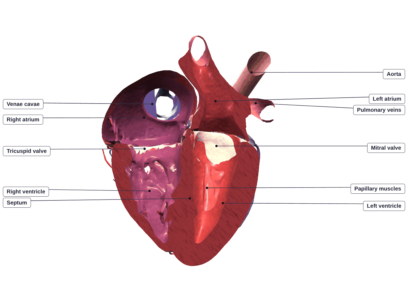
Or press “Follow a blood cell”. Try “Cut open” to see the four chambers and valves.
Heartbeat
View
Find the parts 0 / 14
From outside you see the heart in real colors, with yellow fat in its grooves. Cut open, the walls stay real, with a faint code on the inner lining: a blue tint on the right side (oxygen-poor blood) and a red tint on the left (oxygen-rich blood). Real blood is always red; blue is only a code. Press Real colors to switch to a bold blue/red teaching view. With the heart cut open, small markers show which way blood flows; they are not to scale. The heart beats at real speed; press Slow motion to follow each step (the electrical view and the blood-cell tour slow down on their own). Press Labels to name every visible part.
3D anatomy: Human Reference Atlas (HuBMAP, NIH), VH male heart and blood vasculature, CC BY 4.0. License: CC BY 4.0. Simplified and animated for the web by SciSim.
🎓 For teachers: a 40-minute lesson with this page
- Hook (5 min): project the heart full screen (⛶), press Follow a blood cell and let the class call out the next stop before you press Next.
- Explore (10 min): students find all 14 parts and play Name that part. Ask: why is the left ventricle wall so thick?
- Blood’s route + lub-dub (10 min): trace the double circulation, then solve 5 cases on the pressure and ECG graph.
- Pulse + exercise (10 min): take a pulse (15 s × 4), then compare jogging for an average person and an athlete and calculate cardiac output.
- Exit ticket (5 min): the final quiz. Missions are saved in each student’s browser; finishing all 8 unlocks a printable certificate.
Works in Google Classroom and Teams: just share the link. No sign-up, no ads, works on Chromebooks and phones.
🗺️ Mission 4 · Blood’s Route: the Double Circulation
Oxygen-poor blood is coming back from the body. Tap the parts in the order the blood passes through them — all the way round both loops and back to the body.
⏱️ Mission 5 · Lub-Dub Detective
Each case freezes one moment of a heartbeat. Read the graph: when the left ventricle pressure is above the aorta, the aortic valve is pushed open; when it is below the left atrium, the mitral valve opens. Get 4 of 5 right.
✋ Mission 6 · Take a Pulse
Your pulse is the stretch of an artery each time the left ventricle pushes blood out. Press start and count every beat for 15 seconds (tap the button to help you count), then work out the beats per minute.
Try it on yourself: press two fingers (not your thumb, which has its own pulse) gently on the inside of your wrist below the thumb, or at the side of your neck. A normal resting pulse for teenagers and adults is about 60–100 beats per minute.
🏃 Mission 7 · Exercise Lab
Pick an activity. The graph shows heart rate for 1 minute at rest, 3 minutes of the activity and 5 minutes of recovery, for an average person and a trained athlete. Run at least two activities and solve the calculation.
At rest:
Teaching values for healthy teenagers and young adults; real numbers vary from person to person. A rough maximum heart rate is 220 − age.
✅ Mission 8 · Final Quiz
10 questions from a bank of 20. Get 6 or more to complete the mission.
💡 The Idea, Step by Step
Hold up a fist: that is roughly the size of your heart, and it beats about $100{,}000$ times a day without a rest. It is really two pumps side by side. The right pump sends oxygen-poor blood the short trip to the lungs; the left pump sends oxygen-rich blood the long trip around the whole body. Each pump has a small upper chamber that receives blood (an atrium) and a big lower chamber that pushes it out (a ventricle).
Blood runs in a figure-eight of two loops. In the pulmonary circuit the right ventricle pushes blood to the lungs and it comes back to the left atrium, now loaded with oxygen. In the systemic circuit the left ventricle pushes blood to the body and it comes back to the right atrium, now low on oxygen. Four valves act as one-way doors so blood never flows backward: the tricuspid and mitral valves between atria and ventricles, and the pulmonary and aortic valves at the ventricle exits. The left ventricle wall is thickest because it must generate about $120\ \text{mmHg}$ to drive blood around the body, while the right ventricle needs only about $25\ \text{mmHg}$ for the lungs.
One heartbeat is the cardiac cycle: contraction is systole, relaxation is diastole. The atria contract first, then the ventricles. As the ventricles squeeze, the atrioventricular valves slam shut — the first heart sound, lub ($\text{S}_1$). Pressure builds until it tops the arteries, the semilunar valves open, and blood is ejected. As the ventricles relax and pressure falls, the semilunar valves snap shut — the second sound, dub ($\text{S}_2$). How much blood the heart moves per minute is the cardiac output, $CO = HR \times SV$: at rest about $72 \times 70\ \text{mL} \approx 5\ \text{L/min}$.
Press Follow a blood cell and call out each stop before you press Next — watch it wait at each valve until the valve opens. Then press Cut open, choose Step and compare the 3D valves with the readouts and the moving line on the graph: the QRS spike comes just before the lub, the T wave just before the dub. Switch on Electrical to see the signal leave the SA node, pause at the AV node and race down the septum, and press the Exercise preset to watch cardiac output climb.
📐 How the Heart Works
| Phase | What happens |
|---|---|
| 1. Atrial systole | The atria contract and push the last of the blood through the open atrioventricular (tricuspid and mitral) valves into the ventricles — the atrial kick. The ventricles are now full ($\approx 120\ \text{mL}$ end-diastolic volume). |
| 2. Isovolumetric contraction | The ventricles begin to contract. Pressure rises and the AV valves slam shut — the first heart sound (S1, lub). For an instant all four valves are closed and the volume cannot change. |
| 3. Ventricular ejection | Ventricular pressure tops the pressure in the arteries, the semilunar (pulmonary and aortic) valves open, and blood is ejected — to the lungs from the right, to the body from the left. Left-ventricle pressure peaks near $120\ \text{mmHg}$. |
| 4. Isovolumetric relaxation | The ventricles relax; pressure falls below the arteries and the semilunar valves snap shut — the second heart sound (S2, dub). Again all valves are briefly closed. |
| 5. Ventricular filling (rapid) | Pressure in the ventricles drops below the atria, the AV valves open, and blood that has been collecting in the atria rushes in passively. Most filling happens here, before the atria even contract. |
| 6. Reduced filling (diastasis) | Filling slows as the ventricles near full. Meanwhile the atria keep refilling — the right atrium from the venae cavae, the left atrium from the pulmonary veins — ready for the next atrial systole. |
Order of blood flow through the heart
- Superior & inferior vena cava — oxygen-poor blood returns from the body
- Right atrium
- Tricuspid valve
- Right ventricle
- Pulmonary valve
- Pulmonary artery → lungs (CO₂ out, O₂ in)
- Pulmonary veins — oxygen-rich blood returns
- Left atrium
- Mitral (bicuspid) valve
- Left ventricle
- Aortic valve
- Aorta → body, and back to the venae cavae
A handy memory aid: blood goes through a valve every time it leaves a chamber, and the two atrioventricular valves come before the two semilunar ones on each side — “try pulling my aorta” (tricuspid, pulmonary, mitral, aortic).
Parts of the heart
| Part | Where | Job |
|---|---|---|
| Right atrium | Upper right | Receives oxygen-poor blood from the body (venae cavae); holds the SA node |
| Right ventricle | Lower right, front | Pumps blood to the lungs at about 25 mmHg |
| Left atrium | Upper left, back | Receives oxygen-rich blood from the lungs (pulmonary veins) |
| Left ventricle | Lower left, forms the apex | Pumps blood to the body at about 120 mmHg; thickest wall |
| Septum | Between left and right | Separates the two sides; carries the bundle of His |
| Tricuspid valve | Right atrium → right ventricle | 3 leaflets; stops backflow into the right atrium |
| Mitral (bicuspid) valve | Left atrium → left ventricle | 2 leaflets; stops backflow into the left atrium |
| Pulmonary valve | Right ventricle → pulmonary artery | 3 cusps; stops backflow into the right ventricle |
| Aortic valve | Left ventricle → aorta | 3 cusps; coronary arteries open just above it |
| Chordae tendineae & papillary muscles | Inside the ventricles | Hold the AV valve leaflets so they cannot turn inside out |
| Coronary arteries | On the heart’s surface | Supply the heart muscle itself with oxygen |
The double circulation
Mammals use double circulation: blood passes through the heart twice per lap. The pulmonary circuit (right heart → lungs → left heart) sends deoxygenated blood to pick up oxygen; notice that the pulmonary artery is the one artery that carries deoxygenated blood, and the pulmonary veins the one veins that carry oxygenated blood. The systemic circuit (left heart → body → right heart) delivers oxygen to every tissue. Two loops keep the two kinds of blood separate and let each run at its own pressure — gentle for the delicate lungs, forceful for the far-flung body.
What sets the rhythm
The heartbeat is myogenic: it starts inside the heart itself, not from the brain. The sinoatrial (SA) node in the right atrium is the natural pacemaker; its electrical wave spreads across the atria (making them contract), pauses at the atrioventricular (AV) node, then races down the septum and through the ventricle walls so the ventricles contract from the bottom up. The nervous system and hormones such as adrenaline only speed up or slow down this built-in rhythm — raising heart rate and the force of contraction during exercise.
What the ECG shows
An electrocardiogram (ECG) records the heart’s electrical activity through the skin; the green trace on the graph above shows one beat. The small P wave is the signal spreading across the atria from the SA node, just before they contract. The flat stretch after it (the PR interval, about 0.12–0.20 s) is mostly the pause at the AV node. The tall, narrow QRS complex is the signal racing through the ventricles, just before they contract and the AV valves close (lub). The T wave is the ventricles recovering (repolarising), and it ends around the time the semilunar valves close (dub). The atria recover too, but that small signal is hidden inside the QRS.
🎓 Going Further (AP / A-level / Intro College)
Stroke volume, ejection fraction and Starling’s law
Stroke volume is the difference between the volume in the ventricle at the end of filling and at the end of ejection: $SV = EDV - ESV$. At rest roughly $120 - 50 = 70$ mL. The ejection fraction $EF = SV / EDV \approx 70/120 \approx 58\%$ (healthy range about 55–70%). By the Frank–Starling law, the more the ventricle is stretched by filling, the harder it contracts, so a heart automatically pumps out whatever returns to it — the reason both ventricles stay matched beat by beat.
Cardiac output and blood pressure
$CO = HR \times SV$. Mean arterial pressure is roughly $MAP \approx DBP + \tfrac{1}{3}(SBP - DBP)$; for $120/80$ mmHg that is $80 + 40/3 \approx 93$ mmHg (diastole lasts longer than systole, so the mean sits closer to the lower value). Pressure is also $MAP \approx CO \times TPR$, where $TPR$ is the total resistance of the blood vessels, which is why narrowing the arterioles raises blood pressure.
Why the left ventricle wall is thick
By Laplace’s law the stress in a chamber wall is about $\sigma \approx \dfrac{P\,r}{2h}$ (pressure $P$, radius $r$, wall thickness $h$). The left ventricle works at about five times the pressure of the right, so to keep the wall stress down its wall must be much thicker — about three times the right ventricle’s, which is what the cut-open model shows.
Reading the ECG
On standard ECG paper (25 mm/s) one small square is 0.04 s. The PR interval (atria to ventricles, mostly the AV-node delay) is normally 0.12–0.20 s and the QRS complex is narrower than 0.12 s. Heart rate $\approx 1500 / $ (number of small squares between two R waves); 20 squares means 75 beats per minute.
❓ FAQ
The heart has four chambers: two thin-walled atria on top that receive blood, and two thick-walled ventricles below that pump it out. The right atrium receives deoxygenated blood returning from the body through the venae cavae and passes it down into the right ventricle, which pumps it the short distance to the lungs through the pulmonary artery. The left atrium receives oxygenated blood coming back from the lungs through the pulmonary veins and passes it into the left ventricle, which pumps it at high pressure out through the aorta to the whole body. A muscular wall called the septum separates the two sides, so oxygenated and deoxygenated blood never mix.
Key takeaway: atria receive and ventricles pump; the right side sends deoxygenated blood to the lungs, the left side sends oxygenated blood to the body.Double circulation means blood passes through the heart twice on every full trip around the body, in two separate loops. In the pulmonary circuit the right heart pumps deoxygenated blood to the lungs to load oxygen and unload carbon dioxide, and the oxygenated blood returns to the left heart. In the systemic circuit the left heart pumps that oxygenated blood out to the tissues and the deoxygenated blood returns to the right heart. Mammals and birds need this design because it keeps oxygen-rich and oxygen-poor blood completely separate and lets each circuit run at its own pressure: low for the delicate lungs, high for the body. A single loop would lose too much pressure passing through the lungs to supply an active, warm-blooded animal.
Key takeaway: two loops, pulmonary and systemic, re-pressurize blood between the lungs and the body and stop the two kinds of blood from mixing.The four valves are one-way doors that keep blood flowing forward and stop backflow. The two atrioventricular valves sit between each atrium and ventricle: the tricuspid on the right and the mitral on the left. The two semilunar valves guard the ventricle exits: the pulmonary valve into the pulmonary artery and the aortic valve into the aorta. The lub-dub is the sound of valves snapping shut. The first sound, lub, is the atrioventricular valves closing as the ventricles start to contract, stopping blood from flowing back into the atria. The second sound, dub, is the semilunar valves closing as the ventricles relax, stopping blood from flowing back from the arteries.
Key takeaway: valves enforce one-way flow; lub is the AV valves closing as ventricles contract, dub is the semilunar valves closing as ventricles relax.The cardiac cycle is the repeating sequence of contraction and relaxation in one heartbeat. Systole is contraction, diastole is relaxation. First the atria contract and top off the ventricles. Then the ventricles contract; pressure rises and the AV valves slam shut (lub). With all valves briefly closed, pressure climbs until it exceeds the arteries, the semilunar valves open, and blood is ejected into the pulmonary artery and aorta. The ventricles then relax; pressure falls below the arteries, the semilunar valves close (dub), and with all valves shut the ventricles keep relaxing before the AV valves reopen and they fill again. At rest near 72 beats per minute each cycle lasts about 0.8 seconds.
Key takeaway: the cycle alternates systole and diastole, with the valves opening and closing in a fixed order so blood always moves forward.The left ventricle has a far thicker, more muscular wall because it pumps blood at much higher pressure. It drives the systemic circuit, pushing oxygenated blood all the way around the body against considerable resistance, reaching a peak of roughly 120 millimetres of mercury. The right ventricle only pushes blood the short, low-resistance trip through the lungs, at a peak of about 25 millimetres of mercury, so it needs far less muscle. Crucially, both ventricles eject the same volume per beat, about 70 millilitres; the difference is the pressure they generate, not the amount they pump.
Key takeaway: the left ventricle is thicker because it pumps blood at high pressure around the whole body, while the thinner right ventricle pushes blood gently through the nearby lungs.Cardiac output is the volume of blood the heart pumps each minute, equal to heart rate times stroke volume. Stroke volume is the blood ejected by a ventricle in one beat, about 70 millilitres at rest. At a resting rate near 72 beats per minute, cardiac output is roughly 72 times 70, about 5 litres per minute, close to the body total blood volume. During exercise the muscles demand more oxygen, so cardiac output rises sharply: heart rate climbs, driven by the nervous system and adrenaline, and stroke volume also increases as the heart fills more fully and contracts harder. Together these can lift cardiac output to 20 to 25 litres per minute in a fit adult.
Key takeaway: cardiac output equals heart rate times stroke volume, about 5 litres per minute at rest, rising several fold in exercise as both factors increase.These are the three main vessel types, each suited to its job. Arteries carry blood away from the heart and have thick, elastic, muscular walls to handle the high pressure of each beat; the largest is the aorta. Arteries branch into arterioles and then capillaries, microscopic vessels one cell thick where oxygen, nutrients, and carbon dioxide diffuse between blood and body cells. Capillaries join into venules and veins, which return blood to the heart at low pressure, with one-way valves and squeezing muscles helping it back against gravity. Note that arteries are defined by direction, not oxygen: the pulmonary artery carries deoxygenated blood to the lungs.
Key takeaway: arteries carry blood away from the heart at high pressure, capillaries are thin-walled exchange sites, and veins return blood at low pressure using valves.Oxygen-poor blood from the body enters the right atrium through the superior and inferior venae cavae, passes the tricuspid valve into the right ventricle, and is pumped through the pulmonary valve into the pulmonary artery to the lungs. Oxygen-rich blood returns through the pulmonary veins to the left atrium, passes the mitral valve into the left ventricle, and is pumped through the aortic valve into the aorta and out to the body.
Key takeaway: vena cava, right atrium, tricuspid, right ventricle, pulmonary valve, pulmonary artery, lungs, pulmonary veins, left atrium, mitral, left ventricle, aortic valve, aorta.The P wave is the electrical signal spreading across the atria, just before they contract. The QRS complex is the signal spreading rapidly through the ventricles, just before they contract. The T wave is the ventricles recovering (repolarising) before the next beat. The gap between P and QRS is mainly the deliberate delay at the AV node, which lets the atria finish emptying.
Key takeaway: P = atria activated, QRS = ventricles activated, T = ventricles recovering.The coronary arteries are the heart’s own blood supply. The right and left coronary arteries branch from the aorta just above the aortic valve and run over the surface of the heart; the left splits into the left anterior descending and circumflex arteries. If fatty plaque and a clot block one, the heart muscle it supplies is starved of oxygen and starts to die — a heart attack, or myocardial infarction.
Key takeaway: the heart muscle is fed by coronary arteries, not by the blood inside its chambers.Press the tips of your index and middle fingers gently on the inside of your wrist, just below the base of your thumb (the radial artery), or gently on one side of your neck beside the windpipe (the carotid artery) — never both sides at once. Do not use your thumb, which has its own pulse. Count the beats for 15 seconds and multiply by 4 to get beats per minute. Each beat you feel is the artery stretching as the left ventricle pushes blood out.
Key takeaway: Count for 15 seconds and multiply by 4.For teenagers and adults a normal resting heart rate is about 60 to 100 beats per minute; young children have faster rates. Trained endurance athletes often rest at 40 to 60 beats per minute because exercise makes the heart muscle stronger and the left ventricle larger, so each beat pumps more blood. With a bigger stroke volume, fewer beats give the same cardiac output of about 5 litres per minute. A rough guide to the maximum heart rate is 220 minus your age.
Key takeaway: A stronger heart pumps more per beat, so it needs fewer beats at rest.Yes. The chambers, valves, papillary muscles and great vessels come from the Human Reference Atlas, an open 3D atlas made by the NIH-funded HuBMAP program from the Visible Human male data set (shared under a CC BY 4.0 license). SciSim simplified the meshes for the web and added the colors, the heartbeat, the valve motion and the blood-flow markers, which are teaching animations rather than a medical simulation.
Key takeaway: The shapes are from a real human heart; the motion is a teaching animation.⚠️ Misconceptions & Common Errors